Home > Popular Themes > Human Body
Lungs anatomy, artwork

![]()
Wall Art and Photo Gifts from Science Photo Library
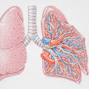
Lungs anatomy, artwork
Lungs anatomy, computer artwork. At top left is the trachea (windpipe, white), which splits into two bronchi, one for each lung (blue). Inside the lungs the bronchi further divide into many bronchioles (small, white). Each bronchiole becomes smaller, finally ending in alveoli (tiny air sacs, bulbous, right), which are the site of gaseous exchange, where oxygen enters the blood and carbon dioxide is removed. Oxygen dissolves in the moist surface of the alveoli and passes into capillaries (red blood vessels) that carry it into the bloodstream. Carbon dioxide passes out of venules (blue blood vessels) into the alveoli and is exhaled through the lungs
Science Photo Library features Science and Medical images including photos and illustrations
Media ID 9220425
© HENNING DALHOFF / SCIENCE PHOTO LIBRARY
Alveolar Alveoli Alveolus Breathing Bronchi Bronchiole Bronchioles Bronchus Capillaries Capillary Chest Deoxygenated Gas Exchange Internal Lung Lungs Organs Oxygenated Oxygenation Physiological Physiology Pulmonary Pulmonology Respiratory System Throat Trachea Veins Venule Venules Vessels Windpipe Blood Vessel Circulation Circulatory System Cutouts Vein
EDITORS COMMENTS
This artwork showcases the intricate anatomy of the lungs, capturing the essence of their vital role in our respiratory system. Against a pristine white background, this computer-generated illustration provides a detailed view of the internal structure and circulation within these essential organs. At the top left corner, we observe the trachea or windpipe, depicted in striking white. From there, it branches into two bronchi, each representing one lung with its mesmerizing blue hue. As we delve deeper into the lungs' intricate network, we encounter numerous bronchioles portrayed as small white pathways. These bronchioles gradually diminish in size until they culminate in alveoli – tiny air sacs that resemble bulbous structures on the right side of the image. The alveoli serve as crucial sites for gaseous exchange where oxygen enters our bloodstream while carbon dioxide is expelled from our bodies. This process occurs through capillaries represented by vibrant red blood vessels that carry oxygenated blood throughout our system. Simultaneously, deoxygenated blood releases carbon dioxide into venules depicted as blue blood vessels which then flows back to be exhaled through our lungs. This visually stunning print not only highlights the complexity and beauty of lung anatomy but also emphasizes their pivotal role in maintaining healthy breathing and sustaining life itself. It serves as a testament to human physiology and invites us to appreciate these remarkable organs responsible for oxygenation within our bodies.
MADE IN THE USA
Safe Shipping with 30 Day Money Back Guarantee
FREE PERSONALISATION*
We are proud to offer a range of customisation features including Personalised Captions, Color Filters and Picture Zoom Tools
SECURE PAYMENTS
We happily accept a wide range of payment options so you can pay for the things you need in the way that is most convenient for you
* Options may vary by product and licensing agreement. Zoomed Pictures can be adjusted in the Cart.